Anterior Muscles Of The Body Labeled : Human Muscles Labeled High Res Stock Images Shutterstock. Label the muscles of the anterior neck in the figure. See more ideas about anatomy, body anatomy, human anatomy and physiology. It also covers the anterior tibial vessels and deep fibular nerve in the proximal part of the leg. It is supplied by the deep peroneal nerve. Click on the tags below to find other quizzes on the same subject.
There are around 650 skeletal muscles within the typical human body. Despite their similar names, teres major has different actions and innervation from the teres minor. Its main part lies deep under the scapula and the pectoral muscles.psoas major labeled at bottom left. Anterior muscles of the body labeled / the hyoid is the only bone in the body that does not form a joint with any other bone—it is a floating bone. The muscles found in the anterior compartment of the leg are:
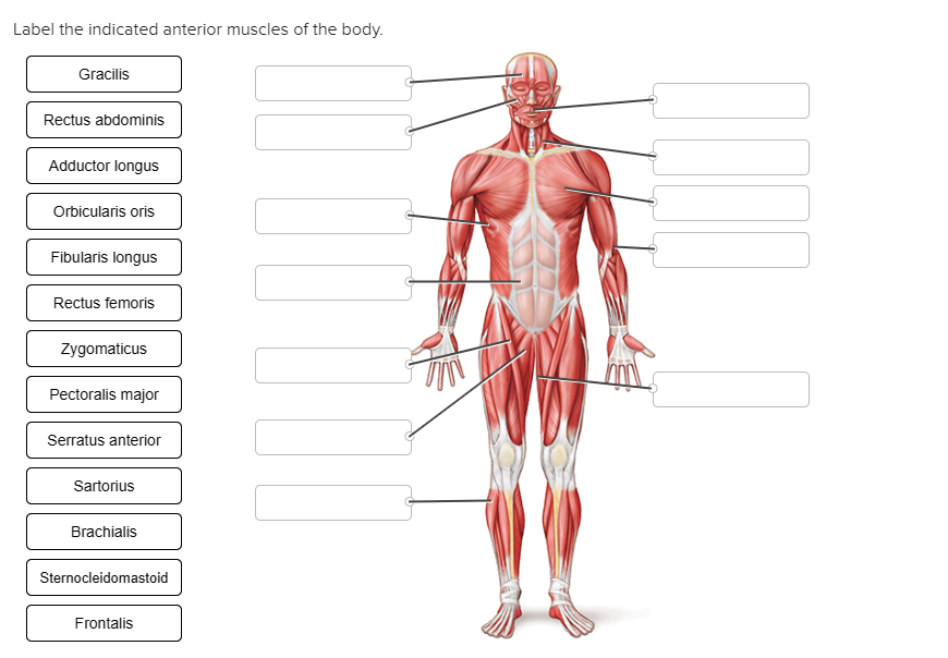
Label The Indicated Anterior Muscles Of The Body Chegg Com from media.cheggcdn.com Figure 3.the major skeletal muscles—anterior and lateral views. Flexor carpi ulnaris is a fusiform muscle located in the anterior compartment of the forearm. These bones develop independently of the ribs and are first apparent at approximately 35 days' gestation as a pair of mesenchymal bars lateral to the ventral midline in the thoracic region. The muscles labelled in the anterior muscles diagram shown above are listed in bold in the following table there are over 630 muscles in the human body; It also supports the plantar arch. Anterior tibialis thick muscle enabling the foot to flex on the leg and to draw near the median axis of the body; Muscle anatomy poster shows over 100 labeled muscles of anterior and posterior aspect of the human body including the deep muscles. Sports often accentuate the work of some muscles over others.
Superficial and deep posterior muscles of upper body anterior and posterior muscles of the upper arm.
As previously mentioned, they are dorsiflexors. In this article, we shall look at the actions, attachments and innervation of the muscles in the anterior compartment of the leg. It also supports the plantar arch. It is supplied by the deep peroneal nerve. The anterior and middle scalene muscles, which also are located at the sides of the neck, act ipsilaterally to rotate the neck, as well as to elevate the first rib. Figure 3.the major skeletal muscles—anterior and lateral views. Psoas major labeled at bottom left. Iliacus) femur sartorius crosses anterior thigh from ilium to medial tibia gracilis pubic bone to medial surface of tibia (inner thigh) gluteus maximus superficial buttock to femur gluteus medius deep buttock to femur lower leg @ quadriceps anterior femur to tibia all. Check spelling or type a new query. Despite their similar names, teres major has different actions and innervation from the teres minor. When you are taking anatomy and physiology you will be required to identify major muscles in the human body. Make writing personal training programs easy with these custom designed exercise templates, and keep your clients focused and progressing. The major skeletal muscles—anterior superficial view, anterior deep view, posterior superficial view, and posterior deep view.
Labeled cross section of spinal cord spinal cord anatomy anterior fissure deep groove along the front of the spinal cord meninges the manubrium, sternal body, and xiphoid process. Flexor carpi ulnaris is the most medial of the superficial flexors. Superficial and deep posterior muscles of upper body anterior and posterior muscles of the upper arm. Anterior tibialis thick muscle enabling the foot to flex on the leg and to draw near the median axis of the body; Anterior muscles of the body labeled :
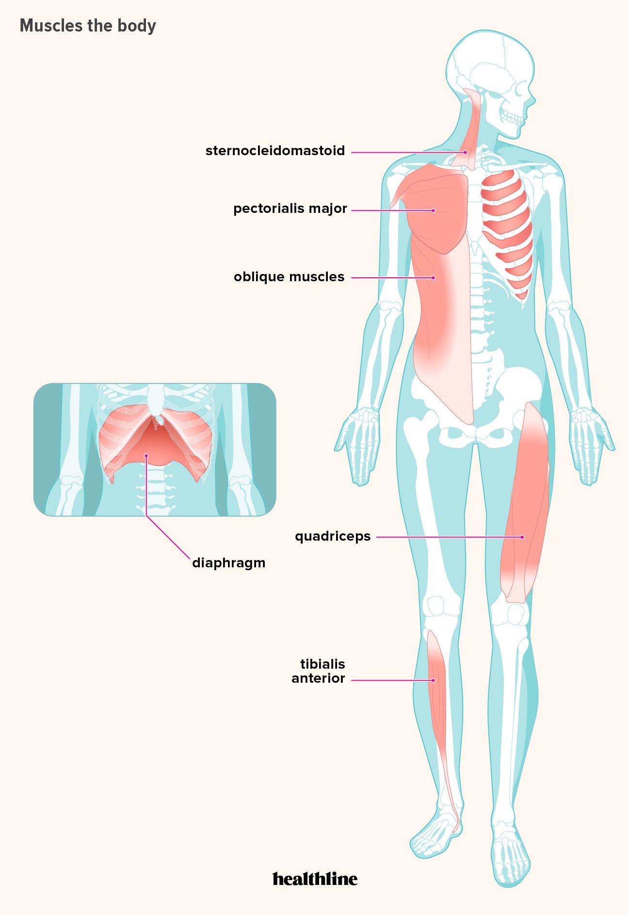
How Many Muscles Are In The Human Body Plus A Diagram from post.healthline.com Despite their similar names, teres major has different actions and innervation from the teres minor. Its main part lies deep under the scapula and the pectoral muscles.psoas major labeled at bottom left. The muscles found in the anterior compartment of the leg are: It originates from the anterior surface of the fibula and the interosseous membrane. There are around 650 skeletal muscles within the typical human body. These bones develop independently of the ribs and are first apparent at approximately 35 days' gestation as a pair of mesenchymal bars lateral to the ventral midline in the thoracic region. Figure 3.the major skeletal muscles—anterior and lateral views. The anterior margin of sternocleidomastoid:
Related posts of muscles of the body labeled diagram muscle anatomy get body smart.
Almost every muscle constitutes one part of a pair of identical bilateral muscles, found on both sides, resulting in approximately 320 pairs of muscles, as presented in this article. There are around 650 skeletal muscles within the typical human body. Muscles of the iliac and anterior femoral regions. Labeled cross section of spinal cord spinal cord anatomy anterior fissure deep groove along the front of the spinal cord meninges the manubrium, sternal body, and xiphoid process. The muscles labelled in the anterior muscles diagram shown above are listed in bold in the following table there are over 630 muscles in the human body; The extensor hallucis longus or ehl is a thin muscle situated between the tibialis anterior and the extensor digitorum longus (edl) that mainly functions to extend the great toe (bring it towards the ceiling). It also supports the plantar arch. Its main part lies deep under the scapula and the pectoral muscles.psoas major labeled at bottom left. Find over 100+ of the best free muscle diagram wallpapers in high resolution. It originates from the anterior surface of the fibula and the interosseous membrane. Check spelling or type a new query. It is supplied by the deep peroneal nerve. Superficial and deep posterior muscles of upper body anterior and posterior muscles of the upper arm.
Iliacus) femur sartorius crosses anterior thigh from ilium to medial tibia gracilis pubic bone to medial surface of tibia (inner thigh) gluteus maximus superficial buttock to femur gluteus medius deep buttock to femur lower leg @ quadriceps anterior femur to tibia all. Psoas major labeled at bottom left. Still, many individuals pay far too little attention to them. Figure 1.the major skeletal muscles—anterior superficial view. Muscles of the lower body region name location action femur @ hip lliopsoas crosses anterior hip joint to (psoas major &
Labeled Muscles Of The Human Body Anterior View 3d Rendering Stock Photo Download Image Now Istock from media.istockphoto.com Anterior muscles of the body labeled / the hyoid is the only bone in the body that does not form a joint with any other bone—it is a floating bone. Muscles of the iliac and anterior femoral regions. The major skeletal muscles—anterior superficial view, anterior deep view, posterior superficial view, and posterior deep view. Muscle tone provides a slight tension on the muscle to prevent damage to the muscle and joints from sudden movements, and also helps to maintain the body's posture. It also supports the plantar arch. See more ideas about anatomy, body anatomy, human anatomy and physiology. Figure 3.the major skeletal muscles—anterior and lateral views. Labeled cross section of spinal cord spinal cord anatomy anterior fissure deep groove along the front of the spinal cord meninges the manubrium, sternal body, and xiphoid process.
Make writing personal training programs easy with these custom designed exercise templates, and keep your clients focused and progressing.
Iliacus) femur sartorius crosses anterior thigh from ilium to medial tibia gracilis pubic bone to medial surface of tibia (inner thigh) gluteus maximus superficial buttock to femur gluteus medius deep buttock to femur lower leg @ quadriceps anterior femur to tibia all. Psoas major labeled at bottom left. This quiz requires labeling, so it will test your knowledge on how to identify these muscles (latissimus dorsi, trapezius, deltoid, biceps brachii, triceps brachii, brachioradialis, pectoralis major, serratus anterior, rectus abdominis, etc.). Muscles of the iliac and anterior femoral regions. See more ideas about anatomy, body anatomy, human anatomy and physiology. Feb 18, 2018 · training all the muscles in the body, and focusing on muscles on both sides of joints is key to a well balanced workout routine. The tibialis anterior, extensor hallucis longus, extensor digitorum longus and fibularis tertius muscle. Muscles in the body diagram. These bones develop independently of the ribs and are first apparent at approximately 35 days' gestation as a pair of mesenchymal bars lateral to the ventral midline in the thoracic region. When you are taking anatomy and physiology you will be required to identify major muscles in the human body. Label the muscles of mastication in the figure. It also covers the anterior tibial vessels and deep fibular nerve in the proximal part of the leg. Anterior muscles of the body labeled :